Первый слайд презентации: Кафедра биологии имени академика В.Н. Ярыгина педиатрического факультета
Parasitic Protozoa. Lumen-Dwelling Protozoa Кафедра биологии имени академика В.Н. Ярыгина педиатрического факультета
Слайд 2: Entamoeba histolytica
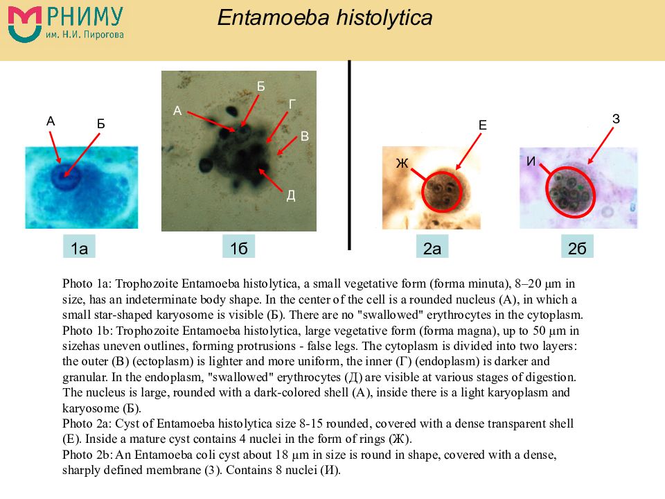
А Б Г В Д А Б Е З Ж И 1а 1б 2б 2а Photo 1a: Trophozoite Entamoeba histolytica, a small vegetative form (forma minuta ), 8–20 µm in size, has an indeterminate body shape. In the center of the cell is a rounded nucleus (A), in which a small star-shaped karyosome is visible ( Б ). There are no "swallowed" erythrocytes in the cytoplasm. Photo 1b: Trophozoite Entamoeba histolytica, large vegetative form (forma magna), up to 50 µm in sizehas uneven outlines, forming protrusions - false legs. The cytoplasm is divided into two layers: the outer ( В ) (ectoplasm) is lighter and more uniform, the inner ( Г ) (endoplasm) is darker and granular. In the endoplasm, "swallowed" erythrocytes ( Д ) are visible at various stages of digestion. The nucleus is large, rounded with a dark-colored shell (A), inside there is a light karyoplasm and karyosome ( Б ). Photo 2a: Cyst of Entamoeba histolytica size 8-15 rounded, covered with a dense transparent shell (E). Inside a mature cyst contains 4 nuclei in the form of rings ( Ж ). Photo 2b: An Entamoeba coli cyst about 18 µm in size is round in shape, covered with a dense, sharply defined membrane (3). Contains 8 nuclei ( И ).
Слайд 3: Balantidium coli
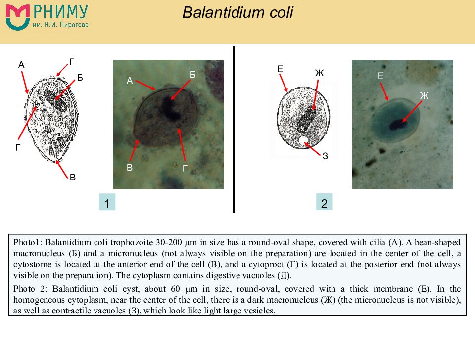
Photo 1: Balantidium coli trophozoite 30-200 µm in size has a round-oval shape, covered with cilia (A). A bean-shaped macronucleus ( Б ) and a micronucleus (not always visible on the preparation) are located in the center of the cell, a cytostome is located at the anterior end of the cell ( В ), and a cytoproct ( Г ) is located at the posterior end (not always visible on the preparation). The cytoplasm contains digestive vacuoles ( Д ). Photo 2: Balantidium coli cyst, about 60 µm in size, round-oval, covered with a thick membrane (E). In the homogeneous cytoplasm, near the center of the cell, there is a dark macronucleus ( Ж ) (the micronucleus is not visible), as well as contractile vacuoles ( З ), which look like light large vesicles. А А Б Б В В Г Г Г Е Е Ж Ж З 1 2
Слайд 4: Lamblia intestinalis = Giardia intestinalis
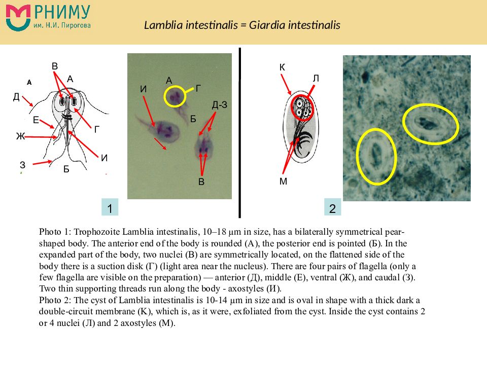
1 2 А Б В А Б В Г Г Д Е Ж З Д - З И И К Л М Photo 1: Trophozoite Lamblia intestinalis, 10–18 µm in size, has a bilaterally symmetrical pear-shaped body. The anterior end of the body is rounded (A), the posterior end is pointed ( Б ). In the expanded part of the body, two nuclei ( В ) are symmetrically located, on the flattened side of the body there is a suction disk ( Г ) (light area near the nucleus). There are four pairs of flagella (only a few flagella are visible on the preparation) — anterior ( Д ), middle ( Е ), ventral ( Ж ), and caudal ( З ). Two thin supporting threads run along the body - axostyles ( И ). Photo 2: The cyst of Lamblia intestinalis is 10-14 µm in size and is oval in shape with a thick dark a double-circuit membrane (K), which is, as it were, exfoliated from the cyst. Inside the cyst contains 2 or 4 nuclei ( Л ) and 2 axostyles (M).
Последний слайд презентации: Кафедра биологии имени академика В.Н. Ярыгина педиатрического факультета: Trichomonas vaginalis
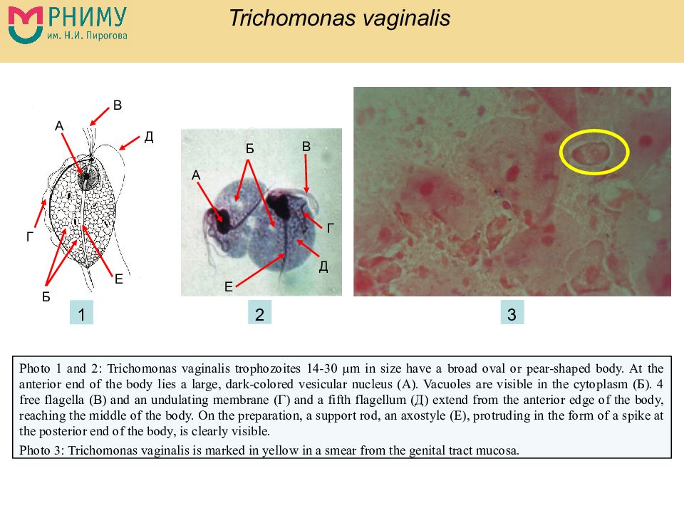
Photo 1 and 2: Trichomonas vaginalis trophozoites 14-30 µm in size have a broad oval or pear-shaped body. At the anterior end of the body lies a large, dark-colored vesicular nucleus (A). Vacuoles are visible in the cytoplasm ( Б ). 4 free flagella ( В ) and an undulating membrane ( Г ) and a fifth flagellum ( Д ) extend from the anterior edge of the body, reaching the middle of the body. On the preparation, a support rod, an axostyle (E), protruding in the form of a spike at the posterior end of the body, is clearly visible. Photo 3: Trichomonas vaginalis is marked in yellow in a smear from the genital tract mucosa. А Б В Г Е Д Б А В Г Д Е 1 3 2
